Diabetes mellitus is a common metabolic disorder characterized by persistent hyperglycemia that eventually leads to serious complications. The prevalence of type 2 diabetes is much higher than that of type 1 and has reached an epidemic proportion where 10.5% of adults (536 million) were supposed to have type 2 diabetes in 20211. Different factors have been implicated in the development of type 2 diabetes, including insulin resistance, impaired insulin secretion, in addition to excessive glucagon secretion2. A long list of risk factors has been identified to be related to the development of type 2 diabetes. These factors include a sedentary lifestyle, lack of physical activity, obesity, genetic factors and ageing. However, strong evidence is available regarding the direct role of the inflammatory process in the pathogenesis of type 2 diabetes, which is aggravated by the same risk factors mentioned above3. The study which is regarded as the cornerstone of the concept of the association between type 2 diabetes and inflammation was conducted in 1993, where the role of tumor necrosis factor- alpha in the development of type 2 diabetes was revealed in an animal study4. Over the following years, an increasing number of clinical studies have further confirmed this correlation between inflammation and type 2 diabetes in humans. This proven correlation has led to expanded attention concerning the use of inflammatory markers to improve risk stratification for diabetes and to target these biomarkers in preventing and managing diabetes mellitus5. Therefore, the development of type 2 diabetes in patients with prediabetes has been thoroughly investigated, where certain inflammatory markers have been studied for their potential role in this process. Many inflammatory biomarkers contribute to the pathogenesis of type 2 diabetes, the most important of which are C-reactive Protein, Tumor Necrosis Factor-alpha, Interleukin-6 (IL-6), Adiponectin, Leptin, Monocyte Chemoattractant Protein-1 and Plasminogen Activator Inhibitor6. IL-6 is a proinflammatory marker that plays a crucial role in the immune response and has been linked to various autoimmune disorders7. It is essentially required in the acute phase response and during the transformation from the acute to the chronic phase of inflammation in addition to many other functions related to the proper inflammatory response8. Subsequent gluconeogenesis, significant hyperglycemia and subsequent hyperinsulinemia have been observed in patients after subcutaneous injection of IL-69. It is thought that IL-6 has a significant impact on the development of insulin resistance by impairing the phosphorylation of insulin receptors, leading to impaired signaling with a consequent high plasma level of insulin10. On the other hand, the state of prediabetes and its correlation with the development of type 2 diabetes have sparked much interest. The American Diabetes Association (ADA) has recently updated its definition of prediabetes. A person is now considered to have prediabetes if he meets at least one of the following criteria:
- Impaired fasting glucose (IFG): A fasting plasma glucose level of 5.6 to 6.9 mmol/L (100 to 125 mg/dL).
- Impaired glucose tolerance (IGT): A 2-hour glucose level of 7.8 to 11.0 mmol/L (140 to 199 mg/dL) after a 75-gram oral glucose tolerance test (OGTT).
- HbA1c level of 39 to 47 mmol/mol (5.7 to 6.4%)11.
Although prediabetes is regarded as the most important risk factor for the development of type 2 diabetes, many other factors, of metabolic concern, have a significant impact on this process. Some of these factors (e.g., weight) are modifiable and can be targeted to avoid or at least delay the progression into diabetes12. Given these findings, the current study aimed to investigate the potential role of serum IL-6 in the progression of prediabetes into type 2 diabetes by comparing its levels among the 3 studied groups; patients with newly onset type 2 diabetes mellitus, those with prediabetes and healthy individuals.
Materials and methods
Subjects
The current cross-sectional study enrolled 159 randomly selected participants who were attending a private clinical laboratory in Baghdad for routine checking during the period between April 2019 and December 2019. They were equally classified according to their medical history and fasting blood glucose (FBG) level into 3 groups: the healthy control group whose FBG level was less than 110 mg/dl, the prediabetes group whose FBG level was 110-125 mg/dl and the diabetes group whose participants met one of the diagnostic criteria of diabetes mellitus. In the first step of the study, written informed consent was obtained from each individual who agreed to participate in the study, followed by taking a full history and performing a full medical examination, in addition to the evaluation of the previous medical records when available. Demographic and anthropometric characteristics of the participants were also recorded.
Methods
For those who met the initial inclusion criteria, 10 ml of whole blood was collected from each participant by a specialized technician. Two millilitres of the blood were used to estimate the complete blood count and HbA1c, while the remaining volume was centrifuged for 10 minutes at 4000 rpm after being kept in a vacuum tube for 15 minutes for full clotting. The collected serum was divided into two fractions and stored in two separate plain tubes. The serum from the first tube was used to evaluate FBG, serum creatinine, serum ALT and AST activity and serum albumin using a Cobas C111 autoanalyzer. For those who still met the inclusion criteria, the second serum-containing tube was stored at -20°C until the day of IL-6 measurement using enzyme-linked immunosorbent assay (ELISA) technique.
The diagnostic criteria of diabetes mellitus
According to the American Diabetes Association, the following criteria were applied for the diagnosis of diabetes mellitus: FBG ≥ 126 mg/dl, 2-hour postprandial blood glucose ≥ 200 mg/dl, HbA1c ≥ 6.5% or random blood glucose ≥ 200 mg/dl in patients with classical symptoms of hyperglycemia13. Patients with any one of these 4 criteria were diagnosed with diabetes mellitus. The clinical presentation was the main factor in confirming type 2 diabetes mellitus; specific tests were used in selected cases.
Exclusion criteria
Individuals with any of the following criteria were excluded from the study: age < 18 years, any current inflammation, any chronic inflammation, use of drugs that might alter the immune system, use of drugs that might alter blood glucose level, patients with type 1 diabetes, pregnancy or patients already diagnosed with type 2 diabetes and receiving treatment of any type.
Ethical statement
The 1964 Helsinki Declaration and its subsequent amendments, as well as equivalent ethical norms, were followed in all procedures performed in the study involving human subjects, whether they were national or institutional research committees.
Statistical analysis
Statistical analyses were performed using SPSS version 26. Descriptive statistics were first performed in the form of means, standard deviations, and the number of participants in each group. Kolmogrov-Samirnov test was used to evaluate the distribution of data. As the data were not distributed normally, Kruskal Wallis, Mann-Whitney and Spearman tests were used to investigate the differences and the correlations between the measured variables. Statistical significance was set at P < 0.05.
Results
Demographic characteristics
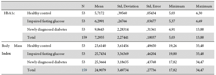
The demographic characteristics of participants are shown in Tables 1 and 2 where sex distribution show a comparable number concerning males and females among the studied groups while BMI distribution shows that obesity is less prevalent among participants compared to the normal or overweight groups. The basal characteristics concerning the measured variables are shown in Table 3.
Comparisons of the studied variables and groups with their correlations
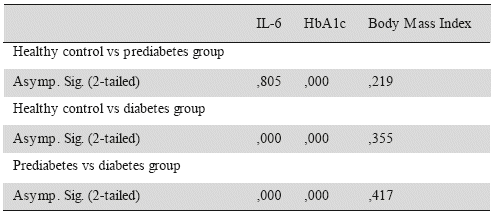
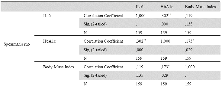
It was evident that there was no significant difference in mean BMI among the 3 studied groups. Using Kruskal Wallis and Mann-Whitney tests, there was no significant difference in mean serum IL-6 between healthy control and impaired fasting glucose groups (P=0.805), while it was significantly higher in the diabetes group compared to both healthy and impaired fasting glucose groups (P=0.00) as shown in Table 4. When compared according to BMI groups, mean serum IL-6 has shown no significant difference among participants with a BMI of < 25 kg/m2, BMI of 25-30 kg/m2 or those with a BMI of > 30 kg/m2 as shown in Table 4. Similarly, no significant difference in mean serum IL-6 has been observed between male and female participants. Spearman’s test was used to determine the strength of correlation between continuous variables. Serum IL-6 has shown a moderate positive correlation with HbA1c (correlation coefficient 0.302) and no correlation with BMI as shown in Table 5.
Discussion
The association between inflammation and the development of type 2 diabetes remains a topic of significant interest in medical studies. Many inflammatory biomarkers have been implicated in the pathophysiology of type 2 diabetes, particularly in subjects with prediabetes whether impaired fasting glucose or impaired glucose tolerance. Persistent attempts have been made to identify single or multiple biomarkers that might predict the possibility of developing type 2 diabetes in individuals with prediabetes. In the current study, it was evident that serum IL-6 is significantly associated with type 2 diabetes, but the question is about whichever was the initial event, did high serum IL-6 precede diabetes mellitus or did this significant increase in serum IL-6 follow the development of diabetes mellitus? Many prospective studies have answered this question and confirmed that IL-6 levels gradually increase in patients with type 2 diabetes in the periods preceding its occurrence. In the current study, we attempted to track serum IL-6 levels during the development of type 2 diabetes. Unexpectedly, subjects in the prediabetes phase showed no significant increase in serum IL-6 compared to healthy subjects, raising doubts about its use as a marker of the transformation of subjects from the prediabetes state to frank diabetes mellitus. Needless to say, this finding does not weaken the concept of the inflammatory role in the development of type 2 diabetes, but it only makes the role of IL-6 questionable. Nevertheless, not being a prospective study might have an impact on the reliability of the results of the current study. Infrequent studies have shown similar findings concerning the difference in serum IL-6 between healthy subjects and those with prediabetes whether impaired fasting glucose or glucose intolerance. The results of the current study were nearly identical to those of a recent study conducted in Indonesia that enrolled 71 participants where plasma IL-6 showed no significant difference between healthy participants and those with prediabetes, in addition to the poor correlation between IL-6 blood glucose levels14. More explicitly, serum IL-6 levels were not significantly different between the healthy control group and subjects with impaired fasting glucose or impaired glucose tolerance but were significantly higher in patients with type 2 diabetes15. More frequent studies have revealed a direct role of serum IL-6 in the development of diabetes mellitus. Certain studies have investigated the molecular level of this correlation and have confirmed that genetic polymorphisms of IL-6 can be used as a biomarker for the early diagnosis of type diabetes16. Surprisingly, serum IL-6 levels were positively correlated with hyperglycemia and possible diabetes mellitus, even in patients with a history of acute pancreatitis17. Even in patients with early autoimmune diabetes mellitus (type 1 and latent autoimmune diabetes of adults) which is characterized by the presence of autoantibodies, specifically anti-islet cells, and their first-degree relatives, it has been shown that serum IL-6 was significantly higher than healthy control group suggesting a potential role of serum IL-6 in the prediction of type 1 diabetes and latent autoimmune diabetes of adults as well18. It is evident that the results concerning the predictive value of IL-6 for the development of type 2 diabetes are inconsistent. Abdominal obesity observed in a significant percentage of participants in various studies is thought to be a major contributor to the increase in serum IL-619. Furthermore, certain studies have shown a significant increase in serum IL-6 that reached 100 folds after an acute exercise20 where the duration and the intensity of the intensity of the exercise were the main determinant factors of serum IL-621. The findings of previous studies suggest a relatively low specificity of serum IL-6 as a predictor of type 2 diabetes, as it seems to be high in other conditions such as type 1 diabetes mellitus and previous acute pancreatitis.


 texto en


